Comparison of muscle activity associated with structual differences in dental hygiene mirrors ~
Division of Dental Hygiene, University of Missouri-Kansas City.
PURPOSE: Ergonomic studies suggest that the commonly used pinch grasp, held in a static position, is a contributing factor for dental Hygienists' development of work-related musculoskeletal disorders (WMSDs) such as carpal tunnel syndrome (CTS), Trigger Thumb, de Quervain's stenosing tenosynovitis, and carpometacarpal (CMC) osteoarthritis. The pinch grasp is commonly used by the dental hygienist while holding the dental mirror in the non-dominant hand. In response to this concern, manufacturers are redesigning dental mirror handles. The value of these re-designed products is based solely on anecdotal evidence. To date, minimal research has been done to examine the non-dominant mirror hand. The purpose of this study was to objectively evaluate dental mirror handle design using surface electromyography (sEMG) to compare muscle activity associated with grasping the mirror. METHODS: This randomized controlled clinical trial utilized a two-by-two repeated measures statistical design. Data was collected on a convenience sample of 19 (N=19) healthy dental hygiene students in their last year of study. Data collection was divided into two phases to maintain a balanced study. The independent variables in phase I were diameter and weight. The independent variables in phase II were weight and padding. Muscle activity was measured while grasping various dental hygiene mirrors in 30-second increments using sEMG. Following data collection subjects designated which mirror felt most and least comfortable to compare subjective data with objective data. RESULTS: Three statistically significant results occurred. In phase II, padding (p=.01) demonstrated the largest reduction of muscle activity in the flexor pollicis brevis, by decreasing mean muscle activity by 3.7 microv. The interaction of diameter and weight (p=.01) in phase I reduced the mean muscle activity in the extensor digitorum by .8 microV and weight (p=.02) in phase II decreased the muscle activity in the extensor digitorum by .62 microV. Self-reports of comfort reported by the subjects in this study were not consistent with the measurements of muscle activity using sEMG. CONCLUSION: Ergonomic adaptations to dental hygiene mirror handles were associated with increases and decreases in muscle activity. The clinical impact of this is amplified as force is exerted. Furthermore, it may be possible to reduce WMSDs for dental hygienists by using instrument designs during the workday. Self-reports of comfort by the subjects in this study did not calibrate with the measurements of muscle activity using sEMG. Additional research is needed to further isolate the external variables of the study and to determine what actual reduction in muscle activity is significant for maintaining musculoskeletal health.
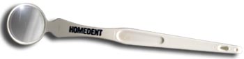
Dental Mirror for inspecting gum tissue and teeth
The DENTAL MIRROR is used to inspect your gum tissue and all the many hidden areas on the backside of teeth. For best results, look into a wall mirror and reflect back into the dental mouth mirror.
USE THE DENTAL MIRROR TO CHECK FOR:
1. Swollen or irritated looking gums.
2. Bleeding gums.
3. Tooth Decay (brown or black spots).
4. Heavy tartar build-up.
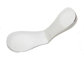 The Full Arch Intraoral Dental Mirror is designed to provide the highest quality digital photo in dentistry. The Full Arch Intraoral Dental Mirror allows you to see a clear, accurate picture of both the maxillary and mandibular arch of your patient's mouth in two simple photos. By capturing a complete and accurate digital photo, the Full Arch Intraoral Dental Mirror will help to present a clear treatment plan, and ease the acceptance process.
Perhaps the most unique feature for the Full Arch Intraoral Dental Mirror is the lightweight, easy to use design. While most dental mirrors only capture half of each arch, our dental mirror is made to capture the full arch of the mouth while sitting comfortably on the the adjacent teeth. The contours made on the dental mirror are to insure your patients full comfort as well as making the photo nearly effortless to capture. Each The Full Arch Intraoral Dental Mirror comes with two distinctly sized lobes at each end. This provides the user an alternative so they can capture a full arch image for most any sized mouth. The stainless steel material is used to help prevent scratching and discoloration during sterilization thus making the dental mirror more durable than other dental products.